Discovering an unexpected dent or indentation in your leg muscle can be concerning, particularly when it appears without obvious explanation. These muscular depressions can manifest for various reasons, ranging from acute traumatic injuries to chronic conditions affecting muscle integrity and contour. Understanding the underlying mechanisms behind muscle indentations is crucial for proper assessment and appropriate medical intervention when necessary.
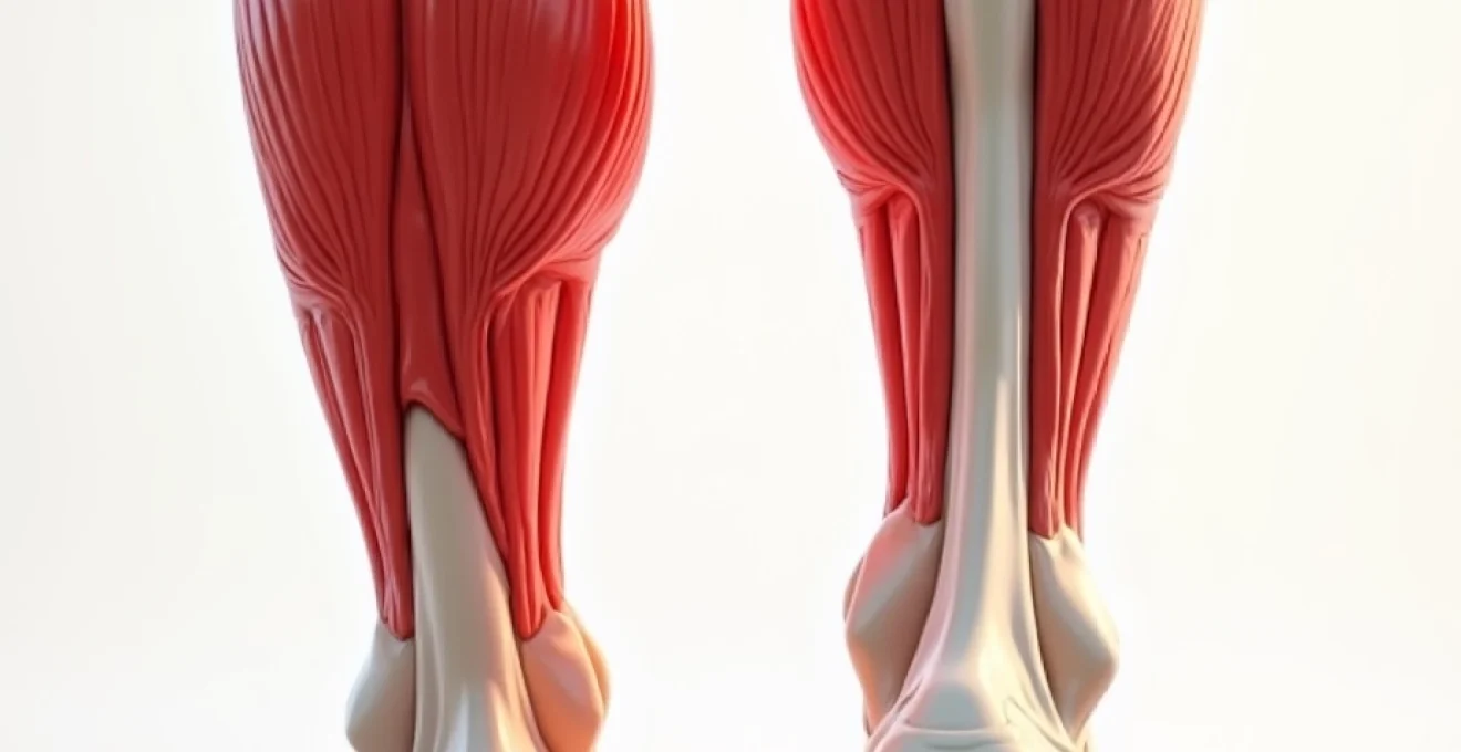
The human leg contains multiple muscle groups working in complex coordination to facilitate movement, stability, and support. When any disruption occurs to this intricate system, whether through direct trauma , vascular compromise, or neurological dysfunction, visible changes to muscle architecture may become apparent. These alterations can present as temporary swelling followed by indentations, permanent muscle atrophy, or localised tissue changes that create noticeable contour abnormalities.
Modern diagnostic techniques have significantly improved our ability to identify and characterise muscle abnormalities in the lower extremities. Healthcare professionals now utilise advanced imaging modalities alongside clinical examination to determine the precise cause of muscular indentations, enabling targeted treatment approaches that address both symptoms and underlying pathology.
Traumatic muscle contusions and haematoma formation in lower extremities
Direct impact injuries to the leg muscles represent one of the most common causes of visible muscle indentations. When external force strikes muscle tissue with sufficient intensity, it can cause immediate tissue damage, bleeding within the muscle fibres, and subsequent swelling that may leave lasting contour changes once the acute inflammatory response subsides.
Direct impact injuries to quadriceps and hamstring muscle groups
The quadriceps and hamstring muscle groups, being prominently positioned and relatively exposed, frequently sustain direct impact injuries during sporting activities, falls, or motor vehicle accidents. These large muscle masses can absorb considerable force before showing visible damage, but when injury does occur, the resulting tissue disruption can be substantial. The quadriceps femoris , consisting of four distinct muscle heads, is particularly susceptible to contusions from frontal impacts, whilst the hamstring complex at the posterior thigh often sustains injury from falls onto hard surfaces.
Following direct trauma, the affected muscle undergoes a predictable sequence of physiological responses. Initial vasoconstriction gives way to vasodilation and increased capillary permeability, allowing blood and inflammatory mediates to accumulate within the injured tissue. This process creates the characteristic swelling and tenderness associated with acute muscle contusions, which may subsequently result in visible indentations as the inflammatory response resolves and damaged tissue is remodelled.
Intramuscular bleeding mechanisms following blunt force trauma
When blunt force trauma occurs to leg muscles, the immediate concern involves the extent of intramuscular bleeding and its potential consequences. Haematoma formation within muscle tissue can create pressure effects that compromise local circulation and lead to secondary tissue damage. The severity of bleeding depends on multiple factors, including the force and direction of impact, the specific muscle involved, and the individual’s underlying vascular health.
Intramuscular haematomas progress through distinct phases of healing, each with characteristic clinical features. During the acute phase, blood accumulation creates a mass effect that may be palpable beneath the skin. As the haematoma organises and begins to resolve, the breakdown products of blood create a visible discolouration pattern that progresses from deep purple-black through various shades of blue, green, and yellow before finally disappearing. The resolution process can leave behind areas of fibrotic tissue that appear as permanent indentations in the muscle contour.
Compartment syndrome risk assessment in tibialis anterior injuries
The anterior compartment of the leg, housing the tibialis anterior muscle along with the extensor digitorum longus and extensor hallucis longus, is particularly vulnerable to compartment syndrome following traumatic injury. This condition occurs when pressure within the fascial compartment rises to levels that compromise tissue perfusion and viability. The relatively inelastic fascial boundaries of the anterior compartment mean that even modest amounts of bleeding or swelling can create dangerous pressure increases.
Acute compartment syndrome represents a surgical emergency requiring immediate fasciotomy to prevent permanent muscle damage and functional loss. However, chronic compartment syndrome can also develop gradually, particularly in athletes engaged in repetitive activities. This condition may manifest as exercise-induced muscle pain, tightness, and eventually, visible changes to muscle contour as chronic pressure effects lead to localised muscle atrophy or fibrosis.
Delayed-onset muscle soreness (DOMS) versus acute contusion differentiation
Distinguishing between delayed-onset muscle soreness and acute muscle contusion can be challenging, particularly when the traumatic event is minor or goes unnoticed. DOMS typically develops 24-72 hours after unaccustomed exercise and is characterised by muscle stiffness, tenderness, and temporary reduction in strength. While DOMS can cause temporary muscle swelling that might create subtle contour changes, these alterations are typically symmetrical and resolve completely within several days.
In contrast, acute muscle contusions usually present with immediate symptoms including localised pain, swelling, and potential visible deformity. The affected area may feel firm or hard to touch, and movement of the injured muscle often exacerbates discomfort. Persistent indentations following what initially appeared to be simple muscle soreness should prompt consideration of underlying tissue damage requiring professional evaluation.
Myofascial trigger points and muscle knot development
Myofascial trigger points represent localised areas of muscle hypercontractility that can create palpable nodules or bands within affected muscles. These areas of dysfunction can develop following trauma, overuse, or postural stress, and may persist long after the initial precipitating factor has resolved. The presence of active trigger points can significantly alter normal muscle function and contribute to the development of visible contour irregularities.
Gastrocnemius and soleus trigger point manifestations
The posterior compartment muscles of the calf, particularly the gastrocnemius and soleus, frequently develop trigger points that can create noticeable changes in muscle contour. These trigger points often manifest as firm, tender nodules that can be palpated through the skin and may create visible indentations in the muscle belly. The gastrocnemius, with its prominent medial and lateral heads, is particularly susceptible to trigger point development following activities involving repetitive plantar flexion or sudden dorsiflexion movements.
Soleus trigger points tend to develop deeper within the muscle and may not be as readily palpable as those in the gastrocnemius. However, they can still contribute to altered muscle function and may create subtle changes in the overall contour of the posterior calf. These trigger points often refer pain to other areas of the leg and can contribute to the development of compensatory movement patterns that place additional stress on other muscle groups.
Piriformis syndrome and deep gluteal muscle adhesions
The piriformis muscle, located deep within the gluteal region, can develop trigger points and adhesions that affect both local muscle function and the function of adjacent muscles in the posterior thigh. Piriformis syndrome occurs when this muscle becomes tight or inflamed, potentially compressing the nearby sciatic nerve and creating symptoms that extend down the entire leg. The resulting muscle dysfunction can lead to compensatory changes in other leg muscles, potentially contributing to the development of visible contour abnormalities.
Deep gluteal muscle adhesions can develop following injury, prolonged sitting, or repetitive activities that place stress on the hip and pelvis. These adhesions can create areas of restricted muscle movement that appear as indentations or irregularities in the normal muscle contour. The complex anatomical relationships within the gluteal region mean that dysfunction in one area often affects multiple muscle groups, potentially creating widespread alterations in lower limb muscle appearance and function.
Iliotibial band syndrome and lateral thigh indentations
The iliotibial band, a thick fascial structure running along the lateral aspect of the thigh, can become tight and inflamed, particularly in runners and cyclists. When ITB syndrome develops, the affected tissue may create visible indentations along the lateral thigh, particularly in the area overlying the vastus lateralis muscle. These indentations may be more prominent during certain movements or muscle contractions, becoming less noticeable when the muscle is relaxed.
The relationship between ITB tightness and underlying muscle function is complex, with restrictions in the fascial tissue often leading to compensatory changes in muscle activation patterns. Over time, these altered movement patterns can contribute to the development of trigger points and adhesions within the vastus lateralis and other lateral thigh muscles, potentially creating permanent changes to muscle contour even after the initial ITB symptoms have resolved.
Vastus lateralis contracture following overuse injuries
The vastus lateralis muscle, forming the lateral portion of the quadriceps group, is particularly susceptible to overuse injuries that can lead to the development of contractures and visible contour changes. These injuries often occur in athletes engaged in activities requiring repetitive knee extension, such as cycling, running, or jumping sports. The resulting muscle contracture can create areas of firm, non-pliable tissue that appear as indentations or irregularities in the normal muscle contour.
Contractures within the vastus lateralis can also develop secondary to prolonged immobilisation or as a complication of previous injuries. The progressive nature of muscle contracture means that early intervention is crucial to prevent permanent changes to muscle architecture. Physical therapy interventions including stretching, manual therapy, and progressive strengthening can help restore normal muscle length and function when implemented appropriately.
Neurological conditions affecting lower limb muscle contours
Neurological disorders can significantly impact muscle mass and contour through various mechanisms including denervation atrophy, altered muscle activation patterns, and changes in muscle tone. These conditions may present with gradual or sudden onset of muscle wasting that creates visible indentations or asymmetries in the affected limb.
Peroneal nerve palsy and anterior compartment atrophy
Peroneal nerve injuries can result from direct trauma, compression, or stretch injuries that compromise nerve function and lead to weakness or paralysis of the muscles responsible for foot dorsiflexion and eversion. The common peroneal nerve is particularly vulnerable to injury as it winds around the fibular head, where it can be damaged by direct impact, prolonged compression, or excessive stretch during certain movements.
When peroneal nerve function is compromised, the muscles of the anterior and lateral compartments of the leg begin to atrophy due to lack of neural stimulation. This process, known as denervation atrophy, typically becomes visible within several weeks of nerve injury and progresses over months if the nerve damage is not repaired. The resulting muscle wasting creates characteristic indentations in the anterior and lateral aspects of the leg, particularly noticeable in the tibialis anterior muscle belly.
Sciatic nerve compression and posterior thigh muscle wasting
Sciatic nerve compression can occur at various levels along the nerve’s course, from the lumbar spine through the pelvis and down the posterior thigh. When compression is severe or prolonged, it can lead to motor weakness and eventual atrophy of the muscles innervated by the affected nerve branches. The hamstring muscles, being among the first to be affected by proximal sciatic nerve lesions, may show early signs of wasting that appear as visible indentations along the posterior thigh.
The pattern of muscle atrophy associated with sciatic nerve compression often provides clues about the level and severity of nerve involvement. Proximal lesions affecting the nerve before it divides into its major branches typically cause widespread muscle wasting throughout the posterior thigh and leg, whilst more distal lesions may create selective patterns of atrophy affecting specific muscle groups. Progressive muscle wasting associated with neurological symptoms requires prompt evaluation to determine the underlying cause and appropriate treatment approach.
L5-S1 radiculopathy impact on calf muscle architecture
Lumbar radiculopathy involving the L5 or S1 nerve roots can significantly impact calf muscle function and appearance. These nerve roots contribute to the innervation of the posterior compartment muscles, including the gastrocnemius, soleus, and deep calf muscles. When nerve root compression occurs, either from disc herniation, spinal stenosis, or other causes, the affected muscles may gradually lose mass and strength.
The progression of muscle atrophy in L5-S1 radiculopathy typically follows a predictable pattern, with the more superficial muscles showing changes first. The gastrocnemius muscle, being relatively superficial and prominent, often shows visible signs of atrophy before deeper muscles are significantly affected. This selective pattern of muscle wasting can create asymmetrical calf contours that become more pronounced over time if the underlying nerve compression is not addressed adequately.
Early recognition of neurologically-mediated muscle wasting is crucial for preventing permanent changes to muscle architecture and optimising functional outcomes through appropriate intervention strategies.
Vascular insufficiency and muscle tissue changes
Vascular compromise can significantly impact muscle health and appearance through multiple mechanisms including ischaemia, chronic inflammation, and altered tissue metabolism. Poor circulation to leg muscles can result from arterial insufficiency, venous congestion, or microvascular dysfunction, each creating characteristic patterns of tissue change that may manifest as visible contour abnormalities.
Chronic arterial insufficiency typically develops gradually and may initially present with subtle changes in muscle function and endurance before progressing to more obvious structural alterations. The affected muscles may lose bulk and definition as chronic hypoxia impairs normal cellular metabolism and protein synthesis. This process can create diffuse muscle atrophy that appears as generalised loss of muscle contour rather than discrete indentations, though localised areas may be more severely affected depending on the pattern of vascular compromise.
Venous insufficiency can contribute to muscle changes through different mechanisms, primarily involving chronic oedema and inflammatory processes. When venous return is impaired, fluid accumulation in muscle tissues can create pressure effects that compromise local circulation and cellular function. Over time, this chronic congestion can lead to fibrotic changes within muscle tissue that alter normal architecture and create visible indentations or irregularities in muscle contour. The relationship between venous insufficiency and muscle health is particularly relevant in older adults and individuals with underlying cardiovascular conditions.
Peripheral arterial disease represents a significant cause of muscle changes in the lower extremities, particularly affecting the calf muscles which rely heavily on adequate circulation during activity. The intermittent claudication associated with arterial insufficiency reflects the inability of compromised circulation to meet the metabolic demands of exercising muscle. Chronic ischaemia can lead to muscle fibre loss and replacement with fibrous tissue, creating permanent changes to muscle contour that may persist even after vascular intervention.
Inflammatory myopathies and autoimmune muscle disorders
Inflammatory conditions affecting muscle tissue can create localised or generalised changes in muscle architecture that manifest as visible contour abnormalities. These conditions range from infectious myositis to autoimmune disorders that specifically target muscle tissue, each with characteristic patterns of presentation and progression that can help guide diagnosis and treatment.
Polymyositis and dermatomyositis represent the most common autoimmune inflammatory myopathies affecting skeletal muscle. These conditions typically cause symmetrical muscle weakness and atrophy, often beginning in the proximal muscle groups before progressing to involve distal muscles. The inflammatory process can create areas of muscle necrosis and subsequent fibrosis that appear as permanent indentations or irregularities in muscle contour. The progression of these conditions can be variable, with some patients experiencing rapid deterioration whilst others have a more indolent course.
Infectious myositis can result from bacterial, viral, or parasitic organisms that directly invade muscle tissue or create secondary inflammatory responses. Bacterial myositis typically presents with acute onset of localised muscle pain, swelling, and systemic signs of infection. The affected muscle may develop areas of necrosis or abscess formation that can leave permanent contour changes even after successful treatment. Viral myositis tends to be more diffuse and usually resolves without permanent sequelae, though some cases may result in persistent muscle weakness or atrophy.
Inclusion body myositis represents a distinct form of inflammatory myopathy that typically affects older adults and has a characteristic pattern of muscle involvement. Unlike other inflammatory myopathies, inclusion body myositis often shows asymmetrical muscle weakness and atrophy, frequently affecting the quadriceps and finger flexor muscles early in the disease course. The selective pattern of muscle involvement can create distinctive contour changes that help differentiate this condition from other forms of myositis.
The recognition of inflammatory myopathies requires careful attention to the pattern and progression of muscle changes, as early intervention can significantly impact long-term functional outcomes and quality of life.
Autoimmune conditions affecting connective tissue, such as systemic lupus erythematosus or systemic sclerosis, can also create secondary effects on muscle tissue that manifest as visible contour
changes. These systemic autoimmune disorders can affect muscle tissue through vascular involvement, inflammatory mediator release, and direct tissue infiltration. The resulting muscle changes may be subtle initially but can progress to create significant alterations in muscle contour and function over time.
Drug-induced myopathies represent another important category of inflammatory muscle disorders that can create visible contour changes. Certain medications, including statins, corticosteroids, and antimalarial drugs, can trigger inflammatory responses within muscle tissue or directly affect muscle metabolism. The pattern of muscle involvement in drug-induced myopathy is often dose-dependent and may resolve with discontinuation of the offending agent, though some cases may result in permanent muscle damage.
Diagnostic imaging techniques for lower extremity muscle abnormalities
Modern diagnostic imaging has revolutionised our ability to evaluate muscle abnormalities in the lower extremities, providing detailed visualisation of muscle architecture, vascular supply, and associated soft tissue changes. The selection of appropriate imaging modalities depends on the suspected underlying pathology, the acuity of presentation, and the specific anatomical structures of interest.
Magnetic resonance imaging represents the gold standard for evaluating muscle tissue abnormalities, offering superior soft tissue contrast and multiplanar imaging capabilities. MRI can differentiate between various types of muscle pathology, including acute haematomas, chronic fibrosis, muscle atrophy, and inflammatory changes. The use of contrast enhancement can provide additional information about tissue vascularity and inflammation, whilst specialised sequences such as diffusion-weighted imaging can help identify areas of acute muscle injury or necrosis.
Ultrasound imaging provides a cost-effective and readily available method for evaluating superficial muscle abnormalities and can be particularly useful for guiding therapeutic interventions such as trigger point injections or aspiration of fluid collections. Real-time ultrasound allows dynamic assessment of muscle function during movement, which can be valuable for identifying subtle functional abnormalities that may not be apparent on static imaging studies. The ability to perform ultrasound at the bedside makes it particularly useful for acute presentations where immediate assessment is required.
Computed tomography, whilst less commonly used for primary muscle evaluation, can provide valuable information about bone involvement and calcifications within muscle tissue. CT imaging is particularly useful when evaluating suspected compartment syndrome or when assessing the extent of muscle involvement in complex trauma cases. The rapid acquisition times associated with modern CT scanners make this modality useful for unstable patients who cannot tolerate longer examination times.
Advanced imaging techniques continue to evolve, with emerging technologies such as elastography and spectroscopy offering new insights into muscle tissue properties and metabolism that may further enhance our diagnostic capabilities.
Electromyography and nerve conduction studies complement imaging findings by providing functional assessment of muscle and nerve activity. These studies can help differentiate between primary muscle disorders and secondary muscle changes resulting from neurological dysfunction. The combination of imaging and electrophysiological studies often provides the most comprehensive evaluation of muscle abnormalities, guiding both diagnosis and treatment planning.
The integration of multiple diagnostic modalities allows healthcare professionals to develop a comprehensive understanding of muscle abnormalities and their underlying causes. What initially appears as a simple muscle indentation may represent a complex interplay of traumatic, vascular, neurological, or inflammatory processes that require targeted intervention to prevent progression and optimise functional outcomes. Early recognition and appropriate evaluation of muscle contour changes can significantly impact treatment success and long-term prognosis for patients experiencing these concerning symptoms.