The human eye undergoes remarkable transformations throughout life, with the optical axis serving as a fundamental parameter that influences vision quality and refractive status. Understanding whether your eye axis changes over time requires examining the complex interplay between anatomical development, ageing processes, and pathological conditions that can alter ocular dimensions. From birth through advanced age, the eye’s structural components continuously adapt and modify, potentially affecting the precise measurements that determine how light focuses on the retina.
Recent advances in optical biometry have revealed that ocular axial length modifications occur more frequently than previously understood. These changes can significantly impact refractive error development, influencing everything from childhood myopia progression to adult presbyopic adjustments. For individuals concerned about vision stability or considering refractive surgery, recognising the dynamic nature of ocular anatomy becomes increasingly important for making informed decisions about long-term eye care.
Anatomical structure and development of the eye’s optical axis
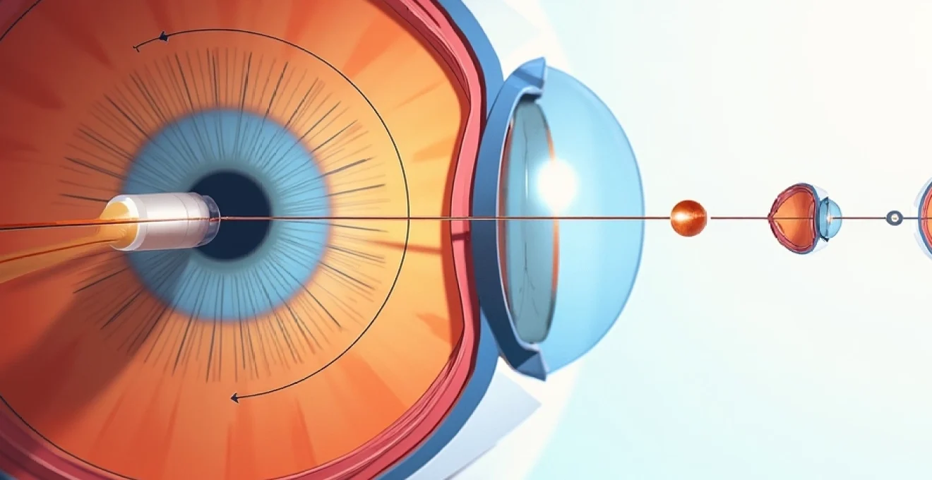
The optical axis of the eye represents an imaginary line connecting the centres of curvature of all refractive surfaces, extending from the anterior corneal surface through the pupil, crystalline lens, and vitreous chamber to reach the retinal fovea. This critical measurement, typically ranging from 22-26 millimetres in healthy adults, determines the eye’s refractive power and focusing ability. Understanding this fundamental structure provides insight into how various factors can influence axial dimensions throughout life.
Corneal curvature changes throughout the human lifespan
Corneal topography undergoes subtle but measurable modifications across different life stages, directly influencing the eye’s optical axis orientation. During infancy and childhood, the cornea exhibits relatively steep curvature that gradually flattens with age. This natural flattening process, known as physiological corneal flattening , typically results in approximately 0.25 to 0.5 dioptres of change over several decades. Environmental factors, including prolonged near work and digital screen exposure, can accelerate these corneal modifications in younger populations.
The corneal astigmatism axis also demonstrates notable stability in most individuals, though significant shifts can occur due to lid pressure, sleeping positions, and mechanical forces applied to the eye during daily activities. Research indicates that against-the-rule astigmatism becomes more prevalent with advancing age, as the vertical corneal meridian flattens more rapidly than the horizontal meridian.
Crystalline lens thickness variations and accommodation effects
The crystalline lens undergoes continuous growth throughout life, adding new lens fibres to its outer cortex while maintaining a relatively stable nucleus. This perpetual growth process results in measurable increases in lens thickness, with studies showing an average annual increase of approximately 0.02 millimetres. These dimensional changes contribute to shifts in the eye’s effective axial length and can influence refractive status, particularly in older adults approaching presbyopia.
Accommodation-related changes also affect the optical axis temporarily during near vision tasks. When focusing on close objects, the lens increases in thickness and curvature, effectively shortening the eye’s focal length. Chronic accommodation demands, particularly in occupations requiring extensive near work, may contribute to permanent structural adaptations in lens geometry over time.
Vitreous chamber elongation in myopic progression
The vitreous chamber represents the largest component of the eye’s axial length, comprising approximately 65% of the total ocular dimension. In myopic eyes, this chamber undergoes progressive elongation, particularly during childhood and adolescence when growth rates are most pronounced. Studies tracking myopic progression reveal that severe myopia can result in axial length increases of 0.3 to 0.5 millimetres annually during peak growth periods.
This elongation process primarily occurs in the posterior segment, where scleral remodelling allows for expansion of the vitreous cavity. The relationship between axial elongation and myopic progression follows a predictable pattern, with each millimetre of axial length increase corresponding to approximately 2.5 to 3.0 dioptres of myopic shift. Understanding this relationship helps predict future refractive changes and guides intervention strategies for myopia management .
Scleral remodeling and posterior staphyloma formation
Scleral tissue undergoes significant remodelling throughout life, particularly in response to mechanical stress and biochemical changes associated with ageing. The sclera’s collagen matrix experiences gradual weakening, making it more susceptible to deformation under intraocular pressure. This process becomes particularly pronounced in highly myopic eyes, where chronic mechanical stress can lead to posterior staphyloma formation.
Posterior staphylomas represent localised outpouchings of the scleral wall that dramatically alter the eye’s posterior contour and effective axial length. These structural changes can develop gradually over decades, creating irregularities in the optical axis that complicate refractive error correction. Advanced imaging techniques now allow clinicians to monitor scleral changes and predict potential complications before significant vision impairment occurs.
Age-related axial length modifications in refractive development
The relationship between age and ocular axial length follows distinct patterns across different life stages, with each period presenting unique characteristics and developmental milestones. Understanding these age-related changes helps explain why refractive errors evolve over time and why some individuals experience significant vision changes while others maintain relatively stable prescriptions throughout their lives.
Childhood emmetropisation process and normal growth patterns
Emmetropisation represents one of nature’s most remarkable self-correcting mechanisms, allowing the developing eye to achieve optimal refractive status through coordinated growth of multiple ocular components. During the first few years of life, the eye undergoes rapid dimensional changes, with axial length increasing from approximately 17 millimetres at birth to nearly 23 millimetres by age six. This growth process occurs alongside corneal flattening and lens power reduction, creating a delicate balance that ideally results in clear distance vision without correction.
The emmetropisation process demonstrates remarkable precision, with healthy children typically achieving refractive errors within ±0.50 dioptres of perfect focus by school age. However, disruptions to this process, whether genetic, environmental, or pathological, can result in persistent refractive errors that continue evolving throughout childhood. Modern lifestyle factors , including increased near work demands and reduced outdoor activities, appear to interfere with normal emmetropisation in an growing number of children.
Adolescent myopic shifts and educational Near-Work impact
Adolescence marks a critical period for refractive development, with many individuals experiencing onset or progression of myopia during secondary school years. The combination of genetic predisposition, intensive educational demands, and reduced outdoor activities creates an environment conducive to axial elongation and myopic progression. Studies indicate that approximately 30% of adolescents develop new-onset myopia during their teenage years, with progression rates varying significantly based on lifestyle factors and genetic background.
Educational systems worldwide report increasing myopia prevalence rates, with some regions experiencing myopia rates exceeding 80% among university-aged students. The relationship between near work intensity and axial elongation becomes particularly pronounced during adolescence, when the eye’s growth mechanisms remain highly responsive to environmental stimuli.
Research suggests that each additional hour of daily near work corresponds to approximately 0.02 dioptres of annual myopic progression during peak adolescent growth periods.
Adult presbyopic changes and lens accommodation decline
Adult eyes typically experience relative stability in axial length measurements, with annual changes rarely exceeding 0.01 millimetres in healthy individuals. However, significant changes occur within the crystalline lens, which continues growing throughout life while gradually losing its accommodative flexibility. The lens increases in thickness and decreases in accommodative amplitude, leading to the inevitable onset of presbyopia around age 40-45 years.
These presbyopic changes affect the eye’s effective focusing system, even though axial length remains relatively stable. The relationship between lens thickness increases and refractive shifts becomes more pronounced with advancing age, contributing to the hyperopic shift commonly observed in older adults. Understanding these changes helps explain why many individuals require reading glasses despite maintaining stable distance vision throughout their adult years. Careful monitoring of these changes allows eye care professionals to anticipate presbyopic onset and recommend appropriate corrective strategies.
Geriatric ocular dimensional stability and pathological variations
Advanced age brings unique challenges to ocular dimensional stability, with pathological conditions becoming increasingly prevalent and influential in determining axial length changes. While healthy ageing eyes typically maintain stable axial dimensions, the development of cataracts, glaucoma, and age-related macular degeneration can significantly alter ocular geometry. Cataract formation, in particular, affects lens thickness and refractive power, sometimes creating dramatic shifts in refractive error that may mask underlying axial length stability.
Geriatric patients also face increased risks of secondary complications from systemic diseases that can affect ocular dimensions. Diabetes mellitus, hypertension, and autoimmune conditions may contribute to structural changes in various ocular tissues, potentially affecting axial measurements. The challenge in this population lies in distinguishing between normal age-related changes and pathological modifications that require intervention. Regular monitoring becomes essential for maintaining optimal vision and detecting potentially sight-threatening conditions in their early stages.
Pathological conditions affecting ocular axial measurements
Various pathological conditions can significantly impact ocular axial measurements, creating changes that extend far beyond normal developmental patterns. These conditions often present complex challenges for both patients and eye care professionals, requiring specialised management approaches and careful monitoring to preserve vision quality. Understanding how different diseases affect axial length provides valuable insights into disease progression and treatment effectiveness.
High myopia and progressive axial elongation syndrome
High myopia, defined as refractive error exceeding -6.00 dioptres or axial length greater than 26 millimetres, represents a pathological condition characterised by progressive axial elongation that can continue throughout adulthood. Unlike physiological myopia, which typically stabilises in early adulthood, pathological myopia demonstrates ongoing progression that can lead to severe complications including retinal detachment, choroidal neovascularisation, and macular hole formation.
The rate of axial elongation in pathological myopia varies considerably among individuals, with some patients experiencing increases of 0.1 to 0.3 millimetres annually well into their adult years. This progressive elongation creates a cascade of structural changes throughout the posterior segment, including scleral thinning, choroidal atrophy, and retinal stretching. Research indicates that eyes with axial lengths exceeding 30 millimetres face dramatically increased risks of vision-threatening complications, making early detection and intervention crucial for preserving long-term visual function.
Studies tracking pathological myopia progression reveal that axial elongation rates correlate strongly with age of onset, genetic factors, and environmental exposures during critical developmental periods.
Understanding these relationships helps guide prevention strategies and treatment protocols for managing progressive myopia in both children and adults.
Keratoconus impact on anterior segment geometry
Keratoconus presents a unique challenge to ocular axial measurements due to its progressive nature and impact on anterior segment geometry. This condition involves gradual thinning and steepening of the central cornea, creating irregular astigmatism and significant changes in corneal curvature that can affect overall axial length calculations. The cone-shaped deformation typically progresses asymmetrically, making accurate biometric measurements increasingly challenging as the condition advances.
Advanced keratoconus can alter the eye’s optical axis orientation, creating complications for both vision correction and surgical planning. The irregular corneal surface disrupts normal light refraction patterns, potentially affecting posterior segment biometry measurements and creating discrepancies in axial length assessments. Modern imaging technologies, including optical coherence tomography and corneal topography, provide detailed maps of corneal irregularities that help clinicians account for these distortions when calculating true axial dimensions.
Diabetic retinopathy and posterior segment structural changes
Diabetic retinopathy introduces complex changes to posterior segment anatomy that can indirectly affect axial length measurements and optical axis stability. Chronic hyperglycemia leads to microvascular changes throughout the retinal circulation, creating areas of ischemia, hemorrhage, and exudation that alter retinal thickness and architecture. Advanced diabetic retinopathy may develop tractional components that physically distort retinal geometry and affect the eye’s optical characteristics.
Diabetic macular edema represents a particularly challenging complication, creating localised changes in retinal thickness that can affect central visual function and biometric measurements. The accumulation of intraretinal fluid alters the normal relationship between anatomical and optical axial length, potentially complicating refractive surgery planning and IOL power calculations. Careful management of diabetic eye disease requires ongoing monitoring of both systemic glucose control and ocular structural changes to minimise progression and preserve visual function.
Glaucomatous optic nerve head excavation effects
Glaucoma creates progressive changes in optic nerve head anatomy that can affect posterior segment measurements and axial length assessments. The characteristic cupping and excavation associated with glaucomatous damage alter the normal contour of the optic disc, potentially affecting the relationship between anatomical landmarks used in biometric calculations. Advanced glaucoma may also involve changes in scleral canal dimensions and peripapillary tissue architecture that influence overall posterior segment geometry.
The relationship between intraocular pressure elevation and axial length changes remains an area of active research, with some studies suggesting that chronic pressure elevation may contribute to subtle axial elongation over time. This relationship becomes particularly relevant in patients with high-pressure glaucoma who experience ongoing structural damage despite treatment. Understanding these connections helps guide treatment strategies and monitoring protocols for preserving both optic nerve function and overall ocular dimensional stability throughout the progression of glaucomatous disease.
Modern biometric technologies for axial length assessment
Contemporary ocular biometry has undergone revolutionary advances with the introduction of sophisticated imaging technologies that provide unprecedented accuracy in axial length measurements. These modern systems utilise various principles including optical interferometry, swept-source optical coherence tomography, and partial coherence interferometry to achieve measurement precision within micrometers. The evolution from traditional ultrasound-based measurements to optical techniques has dramatically improved the reliability and repeatability of axial length assessments across diverse patient populations.
Optical biometry systems such as the IOLMaster 700 and Lenstar LS900 have become gold standards for axial length measurement in clinical practice. These instruments provide comprehensive ocular biometric data including axial length, anterior chamber depth, lens thickness, and corneal curvature measurements in a single, non-contact examination. The superior precision of optical biometry has revealed subtle axial length variations that were previously undetectable with ultrasound techniques, enabling researchers to better understand the dynamic nature of ocular dimensions throughout life.
Advanced swept-source OCT technology represents the latest frontier in axial length assessment, offering enhanced penetration through dense cataracts and providing detailed cross-sectional imaging of the entire ocular axis. These systems generate high-resolution images that allow visualisation of anatomical details throughout the eye’s anterior and posterior segments, facilitating more accurate measurement of individual tissue components contributing to total axial length. Machine learning algorithms integrated into modern biometric devices automatically detect and correct for measurement artifacts, ensuring consistent and reliable results across different patient populations and clinical scenarios.
Clinical implications for refractive surgery planning
Understanding axial length changes over time has profound implications for refractive surgery planning and long-term outcomes. Surgeons must consider not only current refractive status but also anticipated future changes when determining appropriate surgical interventions. For younger patients considering LASIK or PRK, the possibility of continued myopic progression must be carefully evaluated, as additional refractive changes following surgery may necessitate enhancement procedures or alternative correction methods.
Intraocular lens power calculations represent perhaps the most critical application of accurate axial length measurement in modern ophthalmology. The relationship between axial length accuracy and refractive outcomes following cataract surgery follows a predictable pattern, with measurement errors of just 0.1 millimetres potentially resulting in refractive surprises of 0.25 to 0.30 dioptres. This precision requirement becomes even more demanding with premium IOL technologies, where patients expect minimal dependence on spectacles for distance and near vision.
The concept of axial length stability also influences timing decisions for refractive interventions. Patients experiencing active axial elongation may benefit from delaying permanent surgical corrections until dimensional stability is achieved. Conversely, individuals with stable measurements over extended observation periods represent ideal candidates for surgical intervention.
Recent guidelines suggest documenting axial length stability over a minimum 12-month period before proceeding with elective refractive surgery in patients with histories of progressive myopia.
Advanced biometric technologies have also enhanced our understanding of axial length variability in different ethnic populations and geographic regions. Studies utilizing these precise measurement techniques have revealed significant differences in normal axial length ranges across diverse populations, with implications for establishing appropriate normative databases and surgical planning protocols. These population-specific insights help clinicians better predict refractive outcomes and adjust treatment approaches based on individual patient characteristics and demographic factors.
The integration of artificial intelligence and machine learning algorithms into modern biometric systems represents an exciting frontier for axial length assessment. These technologies can identify subtle measurement patterns and predict future changes based on historical data, patient demographics, and risk factors. Predictive modeling capabilities may eventually allow clinicians to anticipate axial length changes years in advance, enabling proactive interventions to prevent myopic progression or optimize surgical timing for better long-term outcomes.
Quality control measures in modern biometric systems have become increasingly sophisticated, incorporating multiple measurement verification protocols and automatic error detection algorithms. These systems can identify measurement inconsistencies, detect patient movement artifacts, and flag potentially unreliable readings before they influence clinical decision-making. The reliability improvements achieved through these technological advances have made axial length monitoring a routine component of comprehensive eye care, enabling earlier detection of pathological changes and more precise surgical planning across all age groups.
Long-term studies utilizing these advanced biometric technologies continue to reveal new insights about axial length stability and change patterns throughout life. Research tracking thousands of patients over decades has identified previously unknown factors that influence ocular dimensional stability, including hormonal changes, systemic medications, and lifestyle modifications. These longitudinal datasets provide invaluable information for counseling patients about expected vision changes and developing personalized monitoring schedules based on individual risk profiles and measurement trends.
The clinical significance of understanding axial length changes extends beyond simple refractive correction to encompass broader aspects of ocular health management. Monitoring axial length changes can provide early warning signs of developing pathological conditions, guide treatment decisions for existing diseases, and help optimize timing for various interventional procedures. Modern eye care increasingly emphasizes the importance of tracking these dimensional changes as part of comprehensive ocular health assessment, recognizing that subtle modifications in axial length often precede clinically apparent vision changes by months or years.
The precision achieved by modern optical biometry has transformed our understanding of ocular dimensional stability, revealing that the eye undergoes continuous subtle changes throughout life that were previously undetectable with traditional measurement techniques.
For patients considering refractive surgery, this enhanced understanding of axial length dynamics has led to more sophisticated pre-operative evaluation protocols and improved long-term outcome predictions. Surgeons now routinely assess not only current axial length measurements but also historical trends and risk factors for future changes when developing treatment plans. This comprehensive approach has significantly reduced the incidence of refractive surprises and improved patient satisfaction with surgical outcomes across all age groups and refractive error types.
The future of axial length assessment promises even greater precision and clinical utility as imaging technologies continue advancing. Emerging techniques such as adaptive optics and ultra-high-resolution OCT may eventually provide cellular-level resolution of ocular structures, enabling detection of microscopic changes that precede measurable axial length modifications. These developments will likely revolutionize our ability to predict and prevent vision-threatening conditions while optimizing refractive outcomes through increasingly personalized treatment approaches tailored to individual patterns of ocular dimensional change throughout life.