The sensation of pressure behind the head and ears represents one of the most challenging diagnostic puzzles in contemporary medicine. This complex symptom constellation affects millions of individuals worldwide, manifesting through a diverse array of anatomical, vascular, and neurological pathways. Understanding the multifaceted origins of retroauricular and occipital pressure requires a comprehensive examination of interconnected physiological systems that influence cranial sensation and pressure regulation.
Modern clinical practice reveals that retroauricular pressure sensations often result from intricate interactions between multiple anatomical structures. The proximity of temporal bone architecture, cranial nerve distributions, and vascular networks creates a complex web of potential symptom origins. Recognition of these interconnected pathways proves essential for accurate diagnosis and effective treatment strategies.
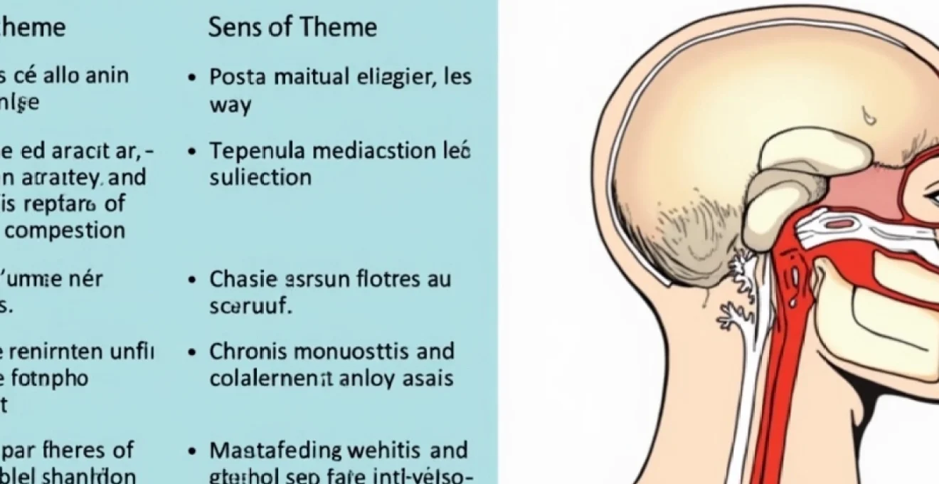
Anatomical structures contributing to retroauricular and occipital pressure sensation
Temporal bone architecture and mastoid process involvement
The temporal bone’s complex architecture plays a fundamental role in retroauricular pressure sensations. The mastoid process, containing numerous air-filled cells connected to the middle ear, creates a potential reservoir for inflammatory processes that can generate significant pressure symptoms. When these mastoid air cells become inflamed or infected, the resulting swelling compresses surrounding tissues and creates the characteristic deep, aching pressure that patients frequently describe.
Anatomical variations in mastoid pneumatisation significantly influence individual susceptibility to pressure-related symptoms. Well-pneumatised mastoid processes provide greater surface area for inflammatory reactions, while poorly pneumatised or sclerotic mastoids may trap secretions more readily. These structural differences explain why some individuals experience more severe retroauricular pressure during upper respiratory infections or allergic reactions.
Trigeminal nerve distribution and auriculotemporal branch pathways
The trigeminal nerve’s auriculotemporal branch provides sensory innervation to the temporal region and external ear, creating direct pathways for pressure transmission. Irritation or inflammation of this nerve branch can produce referred pain patterns that manifest as pressure sensations extending from the temporomandibular joint region towards the ear and posterior head. Dental pathology, particularly involving posterior molars, frequently triggers auriculotemporal nerve symptoms through shared innervation pathways.
Temporomandibular joint dysfunction creates particularly complex pressure patterns through auriculotemporal nerve involvement. Joint inflammation, muscle spasm, or disc displacement can compress the nerve as it passes near the joint capsule. This compression mechanism explains the frequent association between jaw problems and retroauricular pressure complaints.
Occipital nerve anatomy and C2-C3 cervical root origins
The greater and lesser occipital nerves, arising from the C2 and C3 cervical nerve roots, provide crucial sensory innervation to the posterior scalp and occipital region. These nerves traverse complex anatomical pathways, passing through neck musculature before reaching their target territories. Muscle tension, cervical spine dysfunction, or direct nerve irritation can create characteristic occipital pressure sensations that often extend towards the ear region.
Cervical spine pathology, particularly involving the upper cervical segments, frequently generates occipital nerve symptoms through direct root compression or secondary muscle spasm. Facet joint arthritis, disc degeneration, or ligamentous instability can create chronic irritation of these nerve structures. Understanding these anatomical relationships proves essential for developing effective treatment approaches for cervicogenic pressure symptoms.
Eustachian tube dysfunction and middle ear pressure regulation
Eustachian tube dysfunction represents a common yet frequently overlooked cause of retroauricular pressure sensations. The eustachian tube’s role in middle ear pressure equalisation means that dysfunction can create persistent pressure imbalances that manifest as ear fullness, retroauricular discomfort, and associated head pressure. Chronic dysfunction often results from allergic inflammation, anatomical variations, or chronic sinusitis affecting tube patency.
The relationship between eustachian tube function and barometric pressure changes creates particularly troublesome symptoms for sensitive individuals. Weather-related pressure variations can exacerbate underlying dysfunction, creating cyclical patterns of symptom exacerbation. This barometric sensitivity often provides important diagnostic clues for identifying eustachian tube-related pressure symptoms.
Vascular pathophysiology and intracranial pressure mechanisms
Posterior circulation arterial insufficiency and vertebrobasilar system
Vertebrobasilar arterial insufficiency can manifest through posterior head pressure sensations, particularly when combined with positional changes or physical activity. The vertebrobasilar system supplies crucial posterior circulation territories, and compromised flow can create characteristic pressure symptoms accompanied by dizziness, visual disturbances, or balance problems. Age-related vascular changes, atherosclerotic disease, or cervical spine pathology affecting vertebral artery flow contribute to these symptoms.
Subclavian steal syndrome represents an extreme example of posterior circulation compromise, where retrograde flow in the vertebral artery creates characteristic symptom patterns. Exercise-induced arm activity can exacerbate symptoms by increasing subclavian artery demand and reducing vertebrobasilar flow. Recognition of these vascular patterns requires careful attention to symptom timing and associated triggers.
Venous sinus thrombosis and transverse sigmoid junction obstruction
Cerebral venous sinus thrombosis, particularly involving the transverse and sigmoid sinuses, creates characteristic pressure symptoms through impaired venous drainage. The resulting venous congestion increases intracranial pressure and can manifest as severe occipital and retroauricular pressure that worsens with recumbent positioning. Risk factors include hypercoagulable states, dehydration, hormonal contraceptive use, or inflammatory conditions affecting coagulation pathways.
Chronic venous sinus stenosis or idiopathic intracranial hypertension can create similar pressure patterns through sustained elevation of intracranial pressure. These conditions often develop insidiously, with gradual symptom progression that may not immediately suggest vascular pathology. Early recognition proves crucial for preventing permanent neurological complications associated with chronic elevated intracranial pressure.
Idiopathic intracranial hypertension and papilledema correlation
Idiopathic intracranial hypertension presents as elevated intracranial pressure without identifiable underlying pathology, creating characteristic pressure symptoms that often begin in the occipital and retroauricular regions. The condition predominantly affects young women, particularly those with recent weight gain or hormonal changes. Visual symptoms, including transient obscurations or progressive visual field defects, often accompany the pressure sensations.
The relationship between weight gain and idiopathic intracranial hypertension suggests metabolic factors play crucial roles in cerebrospinal fluid dynamics and intracranial pressure regulation.
Papilledema, representing optic nerve head swelling secondary to elevated intracranial pressure, provides objective evidence of pressure elevation and helps distinguish idiopathic intracranial hypertension from other headache disorders. Fundoscopic examination remains essential for evaluating patients with chronic occipital pressure symptoms, particularly when associated with visual complaints or positional variations.
Temporal arteritis and giant cell arteritis manifestations
Giant cell arteritis affecting the temporal arteries creates characteristic pressure and pain patterns in the temporal and retroauricular regions. This inflammatory vasculitis primarily affects individuals over 50 years of age and can present with severe temporal pressure, scalp tenderness, and jaw claudication. The inflammatory process can extend to involve occipital arteries, creating posterior head pressure symptoms.
Early recognition of giant cell arteritis proves crucial for preventing serious complications, including permanent visual loss through ophthalmic artery involvement. Laboratory markers including elevated erythrocyte sedimentation rate and C-reactive protein support the diagnosis, while temporal artery biopsy provides definitive histological confirmation. Prompt corticosteroid treatment can prevent irreversible complications and provides rapid symptom relief.
Infectious and inflammatory aetiologies behind auricular pressure
Acute mastoiditis and coalescent mastoiditis complications
Acute mastoiditis represents a serious complication of middle ear infection, creating intense retroauricular pressure through inflammatory involvement of mastoid air cells. The condition typically develops when middle ear infection extends into the mastoid process, causing characteristic retroauricular swelling, tenderness, and pressure sensations. Progressive bone destruction in coalescent mastoiditis can create life-threatening complications including intracranial extension.
Modern antibiotic therapy has significantly reduced mastoiditis incidence, but the condition remains a serious concern, particularly in immunocompromised individuals or those with antibiotic-resistant organisms. Clinical recognition requires attention to characteristic signs including retroauricular erythema, protrusion of the external ear, and severe pressure symptoms. Prompt surgical intervention may be necessary to prevent intracranial complications or permanent hearing loss.
Chronic otitis media with effusion and mucoid secretions
Chronic otitis media with effusion creates persistent middle ear pressure abnormalities that manifest as retroauricular pressure and fullness sensations. The accumulation of sterile inflammatory fluid behind the tympanic membrane creates pressure imbalances that can extend to involve surrounding structures. Adult presentations often result from eustachian tube dysfunction secondary to allergic rhinitis, chronic sinusitis, or anatomical variations.
The viscous nature of chronic effusions can create particularly troublesome pressure symptoms, as thick secretions resist normal drainage mechanisms. Biofilm formation within chronic effusions further complicates resolution and may require targeted antimicrobial approaches. Addressing underlying eustachian tube dysfunction proves essential for achieving lasting resolution of pressure symptoms.
Petrous apicitis and gradenigo syndrome presentation
Petrous apicitis, representing infection of the petrous apex of the temporal bone, creates a characteristic triad of symptoms including retroauricular pain, sixth cranial nerve palsy, and persistent otorrhoea. This rare but serious condition typically develops as a complication of chronic otitis media or cholesteatoma, with infection extending through pneumatised petrous apex cells.
Gradenigo syndrome, the eponymous term for petrous apicitis, highlights the importance of recognising this condition’s characteristic presentation. The combination of deep retroauricular pressure, diplopia from sixth nerve involvement, and chronic ear drainage should prompt immediate investigation for petrous apex pathology. Advanced imaging studies including high-resolution computed tomography and magnetic resonance imaging prove essential for diagnosis and surgical planning.
Meningitis-related increased intracranial pressure symptoms
Bacterial, viral, or fungal meningitis can create severe occipital and retroauricular pressure through elevated intracranial pressure and inflammatory involvement of pain-sensitive structures. The characteristic triad of fever, neck stiffness, and altered mental status may be accompanied by severe pressure sensations that worsen with movement or changes in position.
Chronic meningitis, particularly fungal or tuberculous forms, can create insidious pressure symptoms that develop gradually over weeks to months. These presentations may lack the acute features of bacterial meningitis, making diagnosis particularly challenging. Cerebrospinal fluid analysis remains the gold standard for diagnosing meningitis and guiding appropriate antimicrobial therapy.
Musculoskeletal origins and cervicogenic headache patterns
Cervicogenic headaches originating from upper cervical spine dysfunction create characteristic pressure patterns that often begin in the occipital region and extend towards the ear. The complex relationship between cervical spine mechanics and cranial pain referral involves both neural and vascular pathways. Upper cervical facet joints, particularly the atlantooccipital and atlantoaxial articulations, possess rich innervation that can refer pain to occipital and retroauricular regions when dysfunction occurs.
Myofascial dysfunction involving suboccipital muscles creates particularly troublesome pressure symptoms through direct compression of occipital nerves and restriction of normal cervical mobility. The suboccipital triangle, bounded by the rectus capitis posterior major and minor and the superior oblique muscles, represents a common site of tension and trigger point development. These muscle groups maintain crucial relationships with occipital nerve pathways, making them frequent contributors to posterior head pressure symptoms.
Postural abnormalities, particularly forward head posture associated with prolonged computer use or mobile device usage, create sustained tension in posterior cervical musculature that can generate chronic pressure symptoms. The biomechanical stress of maintaining abnormal cervical alignment places excessive demands on muscles responsible for head stabilisation. Poor ergonomics and repetitive strain patterns contribute to the development of chronic myofascial pain syndromes that manifest as persistent occipital and retroauricular pressure.
Modern lifestyle factors, including increased screen time and sedentary work environments, have significantly contributed to the rising incidence of cervicogenic pressure symptoms among younger populations.
Whiplash-associated disorders following motor vehicle accidents or sports injuries can create complex patterns of cervical dysfunction that persist long after initial tissue healing. The combination of ligamentous injury, muscle spasm, and joint dysfunction creates multifaceted pressure symptoms that may require comprehensive rehabilitation approaches. Understanding the temporal evolution of whiplash-related symptoms helps distinguish acute injury effects from chronic pain syndrome development.
Neurological conditions manifesting as occipital and retroauricular pressure
Occipital neuralgia represents a distinct neurological condition characterised by sharp, shooting pain and pressure sensations along the distribution of the greater, lesser, or third occipital nerves. The condition can result from nerve entrapment, inflammation, or irritation as these nerves traverse their complex anatomical pathways. Trigger points along the nerve course can precipitate severe pain episodes that patients often describe as electric shock-like sensations accompanied by deep pressure.
Trigeminal neuralgia, while typically associated with facial pain, can occasionally present with atypical patterns that include retroauricular pressure sensations. The close anatomical relationship between trigeminal nerve branches and surrounding structures can create referred pain patterns that extend beyond typical trigeminal territories. Vascular compression of the trigeminal nerve root, most commonly by the superior cerebellar artery, creates the characteristic neurovascular conflict underlying classical trigeminal neuralgia.
Post-herpetic neuralgia following herpes zoster infection of cranial nerve distributions can create persistent pressure and burning sensations in affected regions. The Ramsay Hunt syndrome, involving herpes zoster infection of the geniculate ganglion, can create particularly troublesome retroauricular pressure symptoms accompanied by facial paralysis and vesicular eruptions in the external auditory canal. Early antiviral treatment during acute herpes zoster infection can reduce the risk of developing chronic post-herpetic neuralgia.
Brain tumours, particularly those involving the posterior fossa or cerebellopontine angle, can create pressure symptoms through mass effect and elevated intracranial pressure. Acoustic neuromas, meningiomas, or other space-occupying lesions may present with insidious onset pressure symptoms that gradually worsen over time. Associated symptoms including hearing loss, balance disturbances, or cranial nerve dysfunction provide important diagnostic clues for identifying underlying neoplastic processes.
Chiari malformation, representing herniation of cerebellar tonsils through the foramen magnum, creates characteristic occipital pressure symptoms that worsen with Valsalva manoeuvres or coughing. The malformation can obstruct normal cerebrospinal fluid flow, creating pressure gradients that manifest as posterior head pressure and pain. Magnetic resonance imaging provides definitive diagnosis and allows assessment of cerebrospinal fluid flow dynamics through specialised imaging sequences.
Diagnostic approach and clinical assessment protocols
Comprehensive evaluation of retroauricular and occipital pressure requires systematic assessment of multiple potential contributing factors. The clinical history should explore symptom onset, duration, quality, associated triggers, and relieving factors. Particular attention to temporal patterns, including diurnal variation, relationship to position changes, and association with specific activities, provides crucial diagnostic information for differentiating between various potential causes.
Physical examination must include thorough assessment of the head and neck region, focusing on palpation of tender points, range of motion testing, and neurological evaluation. Examination of the external auditory canal and tympanic membrane can reveal signs of infection or inflammation that might contribute to pressure symptoms. Fundoscopic examination remains essential for detecting papilledema or other signs of elevated intracranial pressure.
Cervical spine assessment should include evaluation of posture, range of motion, and palpation for muscle spasm or trigger points. Provocative testing, including cervical
spine facet loading, upper cervical rotation testing, and assessment for cervical arterial insufficiency help identify musculoskeletal contributions to pressure symptoms. Specialised orthopedic testing can reveal specific joint dysfunction patterns that contribute to cervicogenic pressure manifestations.
Laboratory investigations may be necessary when systemic conditions are suspected. Complete blood count, erythrocyte sedimentation rate, and C-reactive protein levels can identify inflammatory processes such as giant cell arteritis or infectious conditions. Autoimmune markers including antinuclear antibodies may be relevant when connective tissue disorders are considered as potential contributors to pressure symptoms.
Advanced imaging studies play crucial roles in diagnostic evaluation when initial assessment suggests structural abnormalities. High-resolution computed tomography of the temporal bones can reveal mastoiditis, cholesteatoma, or other temporal bone pathology contributing to retroauricular pressure. Magnetic resonance imaging with gadolinium enhancement provides superior soft tissue contrast for evaluating intracranial causes of pressure symptoms, including neoplastic processes, inflammatory conditions, or vascular abnormalities.
Modern neuroimaging techniques, including magnetic resonance angiography and venography, allow non-invasive assessment of cerebrovascular anatomy and can identify vascular causes of pressure symptoms without exposure to ionising radiation.
Specialised diagnostic procedures may be required for specific clinical scenarios. Lumbar puncture with cerebrospinal fluid analysis remains the definitive method for diagnosing meningitis and measuring intracranial pressure. Temporal artery biopsy provides histological confirmation of giant cell arteritis when clinical and laboratory findings suggest this diagnosis. Electrophysiological studies including nerve conduction studies may help localise specific nerve involvement in cases of suspected neuralgia or neuropathy.
Audiological assessment including pure tone audiometry, tympanometry, and acoustic reflex testing can identify hearing-related causes of retroauricular pressure symptoms. These studies help distinguish between conductive and sensorineural hearing loss patterns that might indicate specific pathological processes affecting the ear and surrounding structures. Advanced vestibular testing may be necessary when balance symptoms accompany pressure complaints, particularly in cases of suspected posterior circulation insufficiency or eighth cranial nerve pathology.
The integration of clinical findings with diagnostic test results requires careful consideration of symptom patterns and associated features. Pattern recognition becomes particularly important when multiple potential causes coexist, as is often the case in complex presentations of retroauricular and occipital pressure. Systematic approaches that consider anatomical relationships, temporal patterns, and associated symptoms provide the best framework for accurate diagnosis and effective treatment planning.
Diagnostic challenges arise when symptoms overlap between different conditions or when multiple pathological processes contribute simultaneously to pressure sensations. The development of differential diagnosis algorithms that systematically evaluate potential causes based on clinical presentation, physical findings, and diagnostic test results can improve diagnostic accuracy and reduce unnecessary investigations. Understanding the limitations of various diagnostic modalities helps clinicians make informed decisions about when additional testing is warranted versus when clinical observation and empirical treatment approaches are more appropriate.