When you accidentally slam your finger in a door or drop a heavy object on your toe, the immediate throbbing pain and darkening nail beneath signals a subungual haematoma—blood trapped under the nail plate. This common injury affects millions of people annually, yet many remain uncertain about proper treatment approaches. Understanding the underlying mechanisms of blood accumulation and appropriate therapeutic interventions can mean the difference between rapid healing and prolonged discomfort, potential complications, or permanent nail deformity.
The fingertip represents one of the most densely innervated areas of the human body, making nail bed injuries particularly painful and functionally debilitating. Modern emergency medicine has evolved sophisticated assessment protocols and treatment modalities that can provide immediate relief whilst preventing long-term complications. From simple conservative management to complex surgical interventions, the therapeutic landscape for subungual haematomas requires careful consideration of multiple factors including injury severity, patient age, and underlying nail bed integrity.
Subungual haematoma pathophysiology and blood accumulation mechanisms
The development of a subungual haematoma follows a predictable pathophysiological cascade triggered by direct trauma to the nail unit. When external force strikes the fingertip, the rich vascular network within the nail bed experiences sudden decompression and subsequent capillary rupture. The nail plate, normally adherent to the underlying nail bed through microscopic ridges and grooves, becomes separated by the accumulating blood volume creating a pressurised compartment.
Blood accumulation occurs within the potential space between the nail plate and nail bed, an area that under normal circumstances contains minimal fluid. The rigid nature of the nail plate prevents outward expansion, whilst the firm attachment at the nail fold margins restricts lateral drainage. This anatomical configuration creates a closed compartment where even small volumes of blood can generate significant pressure, explaining why relatively minor injuries can produce disproportionate pain levels.
The temporal dynamics of haematoma formation follow distinct phases beginning with immediate capillary bleeding, followed by platelet aggregation and early clot formation within the first hour. Secondary bleeding may occur over the subsequent 24-48 hours as inflammatory mediators increase local vascular permeability. Understanding these phases becomes crucial when determining optimal timing for therapeutic interventions, as early drainage typically provides superior outcomes compared to delayed treatment approaches.
Biomechanical studies demonstrate that nail plate pressures can exceed 40 mmHg in significant subungual haematomas, substantially higher than normal tissue perfusion pressures. This elevated pressure not only generates intense pain through nociceptor stimulation but can also compromise local circulation, potentially leading to secondary ischaemic damage to the nail matrix and surrounding soft tissues if left untreated for extended periods.
Clinical assessment of fingernail trauma using the hutchinson sign protocol
Comprehensive clinical assessment begins with systematic evaluation of the injured digit, employing established protocols that can differentiate between simple haematomas and more complex nail bed injuries. The Hutchinson sign protocol provides a standardised framework for assessing pigmented lesions beneath the nail, helping distinguish traumatic haematomas from potential malignant processes such as subungual melanoma.
Initial evaluation should document the mechanism of injury, time elapsed since trauma, and patient symptoms including pain intensity and functional limitations. Visual inspection reveals characteristic nail discolouration patterns, typically beginning as bright red coloration that progressively darkens to purple, brown, or black over subsequent hours. The distribution pattern often provides clues about injury severity, with localised haematomas suggesting minor trauma whilst extensive discolouration may indicate significant nail bed laceration.
Nail bed laceration evaluation through transillumination techniques
Transillumination represents a valuable diagnostic technique for evaluating suspected nail bed lacerations beneath intact nail plates. This method involves directing bright light through the fingertip from the palmar aspect, allowing visualisation of underlying anatomical structures through the translucent nail plate. Areas of disrupted nail bed architecture appear as irregular shadows or discontinuities in the otherwise smooth nail bed contour.
The technique proves particularly useful when determining whether nail plate removal is necessary for direct nail bed inspection and repair. Subtle lacerations that might otherwise go undetected can be identified through careful transillumination examination, enabling appropriate treatment planning before undertaking more invasive procedures.
Percentage coverage assessment for subungual blood volume
Quantifying haematoma size through percentage coverage assessment provides objective criteria for treatment decision-making. The widely accepted “50% rule” suggests that haematomas involving greater than 50% of the nail bed surface area warrant aggressive management including potential nail plate removal and direct nail bed repair. Smaller haematomas typically respond well to conservative management or simple drainage procedures.
Accurate percentage assessment requires careful examination under adequate lighting, often enhanced through digital photography with standardised positioning. Some practitioners utilise grid overlay techniques or digital measurement tools to improve assessment accuracy, particularly in medicolegal contexts where precise documentation becomes essential.
Pain scale documentation using the visual analogue score method
Standardised pain assessment through visual analogue scales (VAS) provides objective measurement of patient discomfort levels and treatment effectiveness. The typical 0-10 scale allows patients to quantify their pain experience, with scores above 7 generally indicating severe pain requiring urgent intervention. Serial pain measurements following treatment provide valuable outcome metrics and help identify potential complications.
Throbbing, pulsatile pain characteristic of subungual haematomas often scores highly on standard pain scales, reflecting the pressurised nature of the blood accumulation. Successful drainage typically produces immediate and dramatic pain relief, with VAS scores dropping by 5-7 points within minutes of the procedure.
Differential diagnosis between simple haematoma and nail matrix injury
Distinguishing between isolated subungual haematomas and injuries involving the nail matrix requires careful clinical assessment, as treatment approaches differ significantly. Simple haematomas present with localised blood accumulation beneath an intact nail plate, whilst matrix injuries may demonstrate nail plate disruption, irregular growth patterns, or associated soft tissue lacerations extending beyond the nail fold margins.
Clinical indicators of nail matrix involvement include visible nail plate cracks or splits, particularly those extending to the nail fold, proximal nail bed bleeding, or associated fingertip deformity. These findings often necessitate more complex surgical intervention including nail matrix repair to prevent long-term nail deformity or growth disturbances.
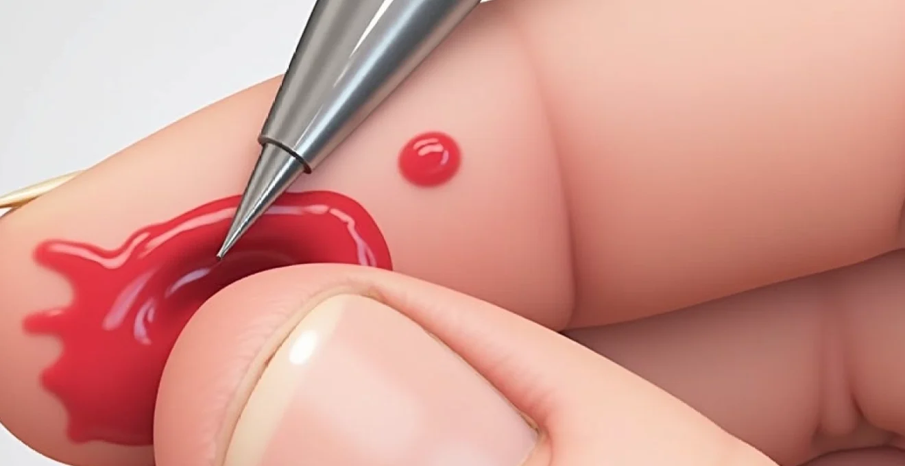
Trephination procedure for subungual haematoma decompression
Trephination represents the gold standard therapeutic intervention for symptomatic subungual haematomas, providing rapid pressure relief through controlled nail plate perforation. This procedure creates a small drainage portal allowing accumulated blood to escape whilst preserving nail plate integrity and avoiding the complications associated with complete nail removal. Success rates exceed 95% when performed within 48 hours of injury, with immediate pain relief reported in the vast majority of cases.
The procedure’s effectiveness stems from basic principles of pressure dynamics and fluid mechanics. Creating even a small aperture in the nail plate dramatically reduces compartment pressure, allowing blood drainage and restoration of normal tissue perfusion. The timing of intervention proves critical, as early drainage prevents clot organisation and maintains liquid blood consistency, facilitating easier evacuation through small drainage holes.
Several trephination techniques exist, each with specific advantages and technical considerations. The choice of method depends on available equipment, practitioner expertise, and patient factors including age, cooperation level, and pain tolerance. All techniques share common principles of aseptic technique, adequate anaesthesia when required, and appropriate post-procedure wound care protocols.
Sterile heated paperclip technique for emergency drainage
The heated paperclip technique represents perhaps the most accessible emergency drainage method, requiring minimal equipment whilst delivering effective results. This approach utilises a standard paperclip heated until red-hot, then applied perpendicular to the nail surface over the point of maximal haematoma accumulation. The intense heat instantly vaporises a small amount of nail keratin, creating a precise drainage hole without mechanical pressure or cutting forces.
Proper execution requires straightening the paperclip to create a stable implement, heating over an open flame or gas burner until glowing, then rapidly applying to the nail surface with gentle pressure. The procedure produces immediate steam as heated metal contacts the nail, accompanied by a characteristic burning keratin odour. Drainage typically begins within seconds of hole creation, often producing dramatic pressure relief.
18-gauge needle perforation method under local anaesthesia
The needle perforation technique offers precise control and reduced patient discomfort through local anaesthetic administration. An 18-gauge needle provides optimal diameter for effective drainage whilst minimising nail plate trauma. The procedure begins with topical anaesthetic application or digital nerve block, followed by careful needle positioning perpendicular to the nail surface.
Gentle rotational pressure gradually advances the needle through the nail plate, with tactile feedback indicating breakthrough into the subungual space. The characteristic “give” sensation confirms successful perforation, typically followed by immediate blood drainage. This technique proves particularly suitable for anxious patients or those requiring multiple drainage attempts due to clot reformation.
Electrocautery trephination using hyfrecator equipment
Electrocautery devices such as the Hyfrecator provide controlled thermal energy for precise nail plate perforation with excellent haemostatic properties. The fine electrode tip allows accurate positioning over the optimal drainage point, whilst adjustable power settings accommodate different nail thicknesses and patient tolerances. This technique combines the precision of needle perforation with the thermal advantages of heated implement methods.
The electrocautery approach produces minimal mechanical trauma to surrounding nail structures whilst creating clean, sealed drainage holes that resist premature closure. Steam evacuation during the procedure indicates successful penetration, followed by rapid blood drainage and pressure relief. Post-procedure bleeding rarely occurs due to the cauterising effect on small blood vessels.
Post-trephination wound care and antiseptic application protocols
Appropriate post-trephination care significantly influences healing outcomes and infection prevention. Initial drainage often continues for 24-48 hours, requiring absorbent dressing materials and regular monitoring for signs of complications. The drainage hole typically remains patent for several days before spontaneous closure through normal nail growth processes.
Antiseptic application protocols recommend twice-daily cleansing with dilute povidone-iodine or chlorhexidine solutions, avoiding alcohol-based preparations that may cause excessive tissue desiccation. Protective dressing should allow drainage whilst preventing external contamination, typically achieved through non-adherent gauze pads secured with adhesive tape avoiding circumferential wrapping that might impede circulation.
The key to successful subungual haematoma management lies in early recognition and prompt intervention, with trephination providing immediate relief when performed within the optimal therapeutic window.
Conservative management strategies for minimal subungual bleeding
Conservative management represents the appropriate first-line approach for minor subungual haematomas involving less than 25% of the nail bed surface area, particularly when patient pain levels remain tolerable and functional limitations are minimal. This strategy relies on natural reabsorption processes to gradually eliminate trapped blood whilst supporting healing through optimal wound care conditions and symptom management protocols.
The rationale for conservative management stems from understanding natural healing mechanisms and the body’s capacity to reabsorb small blood collections over time. Minor haematomas typically undergo predictable resolution phases including initial clot formation, gradual liquefaction through fibrinolytic processes, and eventual reabsorption through lymphatic drainage. This process typically requires 2-4 weeks for complete resolution, during which nail discolouration gradually fades and normal appearance returns.
Initial management focuses on minimising further trauma through protective splinting or padding, whilst addressing pain and swelling through appropriate pharmacological interventions. Elevation of the affected extremity helps reduce venous congestion and promotes lymphatic drainage, potentially accelerating reabsorption processes. Cold therapy application during the first 24-48 hours can limit ongoing bleeding and reduce inflammatory responses.
Patient education plays a crucial role in conservative management success, ensuring individuals understand normal healing timelines and recognise warning signs requiring medical attention. Expectations should be realistic regarding appearance changes, as nail discolouration may persist for months until complete nail plate replacement occurs through normal growth processes. Regular follow-up assessments allow monitoring of healing progress and early identification of complications requiring intervention escalation.
Activity modification recommendations include avoiding repetitive trauma or pressure to the affected digit, temporarily discontinuing activities that might worsen the injury, and maintaining optimal hygiene to prevent secondary infection. Simple analgesics such as paracetamol or ibuprofen typically provide adequate pain control, whilst topical anaesthetic preparations may offer additional symptom relief in some cases.
Surgical intervention criteria and nail bed repair techniques
Surgical intervention becomes necessary when clinical assessment reveals significant nail bed injury extending beyond simple haematoma formation. Complex injuries involving nail bed lacerations, nail matrix damage, or associated fractures require systematic surgical approach to restore normal anatomy and prevent long-term complications such as nail deformity, chronic pain, or functional impairment.
The decision to pursue surgical management depends on multiple factors including injury mechanism, haematoma size and distribution, presence of nail plate disruption, and associated soft tissue damage. High-energy trauma mechanisms such as crush injuries or power tool accidents carry higher risks of complex injury patterns requiring surgical exploration and repair. Patient factors including age, occupation, and functional requirements also influence treatment decisions.
Surgical exploration typically requires adequate anaesthesia, usually achieved through digital nerve blocks using lidocaine with or without epinephrine depending on vascular considerations. The procedure begins with careful nail plate removal when indicated, followed by thorough irrigation and debridement of the nail bed to assess injury extent and remove devitalised tissue or foreign material.
Complete nail avulsion indications using the 50% rule
The 50% rule provides evidence-based criteria for determining when complete nail avulsion becomes necessary for optimal patient outcomes. Haematomas involving greater than 50% of the nail bed surface area frequently indicate underlying nail bed lacerations requiring direct visualisation and repair. This threshold reflects the relationship between haematoma size and likelihood of significant underlying injury requiring surgical intervention.
Complete nail avulsion involves careful separation of the nail plate from the underlying nail bed, typically achieved through gentle traction combined with blunt dissection using appropriate instruments. The procedure requires patience and precision to avoid iatrogenic trauma to the delicate nail bed tissues. Proximal nail fold manipulation may be necessary to access the nail matrix region for complete removal.
Nail bed suturing with 6-0 absorbable chromic catgut
Nail bed repair utilises fine absorbable sutures to restore anatomical continuity and promote optimal healing conditions. 6-0 chromic catgut represents the preferred suture material due to its biocompatibility, appropriate tensile strength, and predictable absorption timeline coinciding with tissue healing phases. The fine diameter minimises tissue trauma whilst providing adequate wound edge approximation.
Precise suture placement proves critical for preventing irregular nail growth patterns or surface irregularities in the regenerated nail plate. Sutures should achieve accurate edge alignment without excessive tension that might compromise tissue perfusion. Interrupted suture patterns typically provide superior results compared to continuous techniques, allowing individual tension adjustment and reducing risks of suture line ischaemia.
Nail plate replacement as biological splint application
The original nail plate, when suitable, can be reimplanted as a biological splint to maintain nail fold anatomy and protect the repaired nail bed during initial healing phases. This technique prevents nail fold adhesions that might interfere with normal nail growth patterns whilst providing natural protection for delicate repair sites. The reimplanted nail plate typically loosens and separates spontaneously as the new nail grows from the matrix.
Nail plate preparation involves thorough cleansing and trimming of irregular edges that might cause trauma during reimplantation. Small drainage holes may be created to prevent fluid accumulation beneath the plate, whilst maintaining sufficient structural integrity to serve as an effective splint. Secure fixation requires careful suture placement avoiding damage to the underlying repair.
Silastic sheet alternative for nail fold preservation
When the original nail plate cannot be salvaged due to extensive damage or contamination, silastic sheeting provides an effective alternative for nail fold preservation and protection of repair sites. Medical-grade silicone sheets offer biocompatibility, appropriate flexibility, and transparency allowing visual monitoring of healing progress. The material can be trimmed to fit individual nail bed dimensions and secured using fine sutures or tissue adhesives.
Silastic sheet placement requires careful positioning to maintain natural nail fold contours whilst avoiding excessive pressure on repair sites. The material should extend slightly beyond the nail bed margins to ensure complete coverage, with smooth edges preventing tissue irritation or trauma. Regular monitoring allows assessment of healing progress and early identification of complications requiring intervention.
Successful nail bed repair requires meticulous attention to anatomical detail
and functional restoration, with meticulous suture placement serving as the foundation for optimal long-term outcomes.
Infection prevention and antibiotic prophylaxis protocols
Preventing secondary infection represents a critical component of subungual haematoma management, as the nail bed’s rich vascular supply and potential communication with deeper structures creates favourable conditions for bacterial proliferation. Contaminated injuries particularly those involving dirty implements or environments, carry significantly elevated infection risks requiring aggressive prophylactic measures and enhanced surveillance protocols.
Standard infection prevention begins with thorough wound irrigation using sterile saline or dilute antiseptic solutions, removing debris, blood clots, and potential bacterial contaminants. High-pressure irrigation proves particularly effective for crushing injuries where particulate matter may embed within nail bed tissues. The irrigation process should continue until return fluid runs clear, indicating adequate contamination removal.
Antibiotic prophylaxis recommendations vary based on injury severity, contamination level, and patient risk factors including diabetes mellitus, immunocompromise, or peripheral vascular disease. Clean injuries in healthy individuals typically require only topical antiseptic application, whilst contaminated wounds or high-risk patients benefit from systemic antibiotic coverage targeting common skin pathogens including Staphylococcus aureus and Streptococcus species.
Current evidence supports short-course antibiotic prophylaxis using first-generation cephalosporins such as cefalexin 500mg twice daily for 5-7 days, providing effective coverage against likely pathogens whilst minimising antibiotic resistance concerns. Alternative regimens include amoxicillin-clavulanate for enhanced anaerobic coverage in severely contaminated wounds, particularly those involving organic matter or soil exposure.
Patient education regarding infection warning signs enables early recognition of complications requiring urgent medical attention. Key indicators include increasing pain beyond expected levels, progressive swelling or erythema extending beyond the immediate injury site, purulent discharge, fever, or red streaking suggesting ascending lymphangitis. Prompt recognition and treatment of developing infections prevents progression to deeper structures including bone or tendon sheaths.
Tetanus prophylaxis requires assessment of immunisation status and wound characteristics, with contaminated injuries typically warranting tetanus toxoid administration regardless of recent vaccination history. Patients with uncertain immunisation status or wounds involving significant contamination may require tetanus immunoglobulin in addition to active immunisation to ensure adequate protection.
Follow-up protocols should include assessment at 48-72 hours post-injury for infection surveillance, wound healing evaluation, and patient education reinforcement. Digital photography can provide objective documentation of healing progress and facilitate remote consultation when necessary. Any deviation from expected healing patterns warrants immediate re-evaluation and potential treatment modification.
Chronic infection complications, though rare, can result in permanent nail deformity, chronic pain syndromes, or functional impairment requiring complex reconstructive procedures. Prevention through appropriate initial management represents the most effective strategy, emphasising the importance of thorough assessment, adequate debridement, and appropriate antibiotic selection when indicated. Understanding these principles enables healthcare providers to deliver evidence-based care whilst minimising long-term complications and optimising patient outcomes.